Key Features
Close-to-natural environmental conditions for sensitive samples
The NEW ready-to-use TruLive3D Dishes facilitate sample mounting
Compact, vibration-free and robust design
Resolution down to 255 nm in xy
Intuitive User Interface
Product Overview
The TruLive3D Imager is optimized for fast 3D multi-sample imaging of delicate live specimens in their native 3D environment.
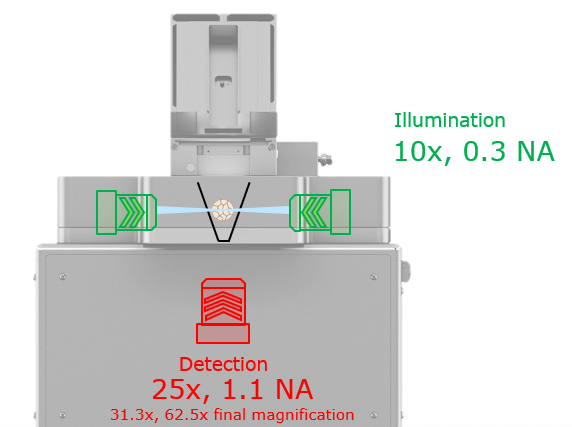
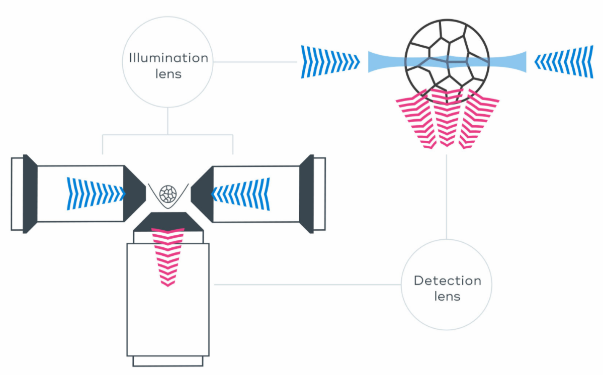
The optical concept, with dual-sided illumination and single-lens detection from below, enables fast acquisition speed, high-resolution imaging, and minimal shadowing effects.
The system is especially suited for multi-position imaging of small embryos (e.g. mouse), 3D spheroids, oocytes and more, enabling time-lapse, high-throughput imaging experiments.

Large Sample Holder
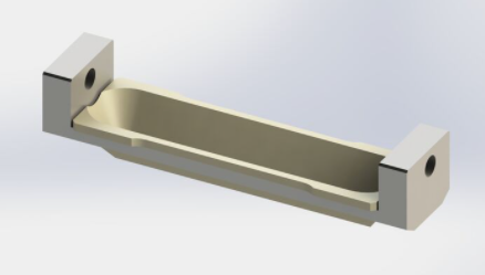
The sample chamber in the TruLive3D Imager fits a large sample holder (length = 75 mm), which can accommodate tens to hundreds of samples.
Similar to the InVi SPIM, the FEP foil lining the holder serves the dual function of a “curved coverglass” and a physical barrier between the immersion medium and sample medium.
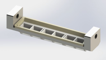
The new ready-to-use TruLive3D Dishes are fully compatible with the TruLive3D Imager. The possibility to line up three disposable dishes extends the capacities of the system to enable the parallel imaging of samples grown under different media conditions.


Device Design
The TruLive3D Imager compact, robust and vibration-free design provides maximal stability even during long-term high-throughput experiments.
Tailored to fit your lab bench, this class 1 laser system does not require any air table or vibration-compensation mechanism as all moving components are light-weight and balanced. Maximal stability of focus and thermal conditions are also guaranteed. The proprietary piezo-crawler stages ensure longevity and precision for a permanently accurate specimen positioning. Neither the images nor the quality of your sample is affected thanks to the unique and very gentle image-acquisition concept.
Illumination and Detection
The TruLive3D Imager can achieve a resolution down to 255 nm in xy, enabling resolving subcellular structures in living samples free of phototoxic effects.
The system features dual-sided illumination and single-lens detection from below, enabling high-resolution imaging and minimal shadowing effects. A wide-field imaging option facilitates sample positioning.
Two Nikon CFI Plan Fluor 10x W 0.3 NA water immersion objective lenses project the light-sheet on the sample. Detection includes a high numerical aperture Nikon CFI Apo 25x W 1.1 NA water immersion objective lens. An additional magnification changer results in 31.3x and 62.5x total. magnification for field view and sampling adjustment according to your experimental needs.
Illumination | Detection | Effective Magnification | Field of View | Pixel Size | Optical Resolution |
10x/0.3NA | Nikon 25x/1.1NA | 31.3x | 420 µm | 208 nm | 255 nm |
62.5x | 210 µm | 104 nm |

| 제품소재 | 상품페이지 참고 |
|---|---|
| 색상 | 상품페이지 참고 |
| 치수 | 상품페이지 참고 |
| 제조자 | 상품페이지 참고 |
| 세탁방법 및 취급시 주의사항 | 상품페이지 참고 |
| 제조연월 | 상품페이지 참고 |
| 품질보증기준 | 상품페이지 참고 |
| A/S 책임자와 전화번호 | 상품페이지 참고 |
사용후기가 없습니다.
상품문의가 없습니다.
등록된 상품이 없습니다.